
Does a Diagnostic X-ray Make You Radioactive?
Ordinary diagnostic X-rays do not leave a patient radioactive. Learn why X-ray imaging and nuclear medicine have different sources and different follow-up questions.
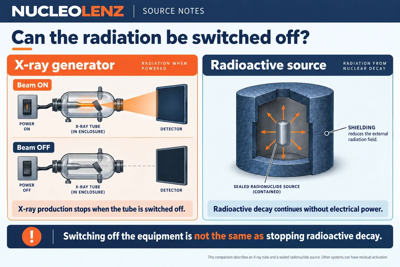
Open the concept graphic at full size ↗
Three key ideas
Where does the radiation come from?
- Diagnostic X-ray: An external machine produces the beam during exposure.
- Nuclear medicine: An administered radiopharmaceutical emits radiation.
- After the procedure: Follow the clinical team’s procedure-specific instructions.
At a glance
Compare the key distinctions
| Focus | What it establishes or needs | Important limit or evidence |
|---|---|---|
| Conventional radiography | External X-ray tube | No radioactive tracer introduced by the X-ray itself |
| CT | External X-ray source | Uses X-rays; not a radioactive tracer by itself |
| Nuclear medicine | Administered radiopharmaceutical | Aftercare depends on the procedure |
| PET/CT | Tracer plus CT X-rays | Identify both parts of the combined examination |
This comparison explains radiation sources; examination and aftercare decisions belong to the clinical team.
The word “radiation” describes several different medical technologies. Understanding how the radiation is produced helps explain why a diagnostic X-ray examination and a nuclear medicine procedure have different follow-up questions.
Exposure is not the same as receiving radioactive material
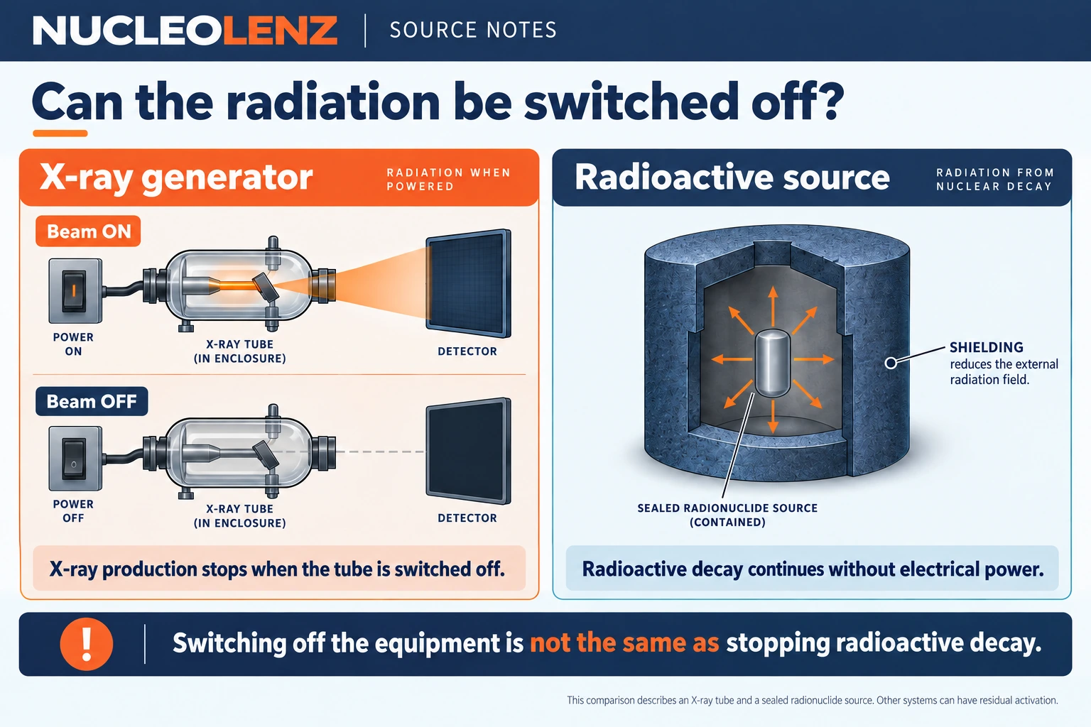
During conventional diagnostic radiography, a machine produces X-rays for the examination. The patient is exposed to those photons; the procedure does not introduce a radioactive tracer. Ordinary diagnostic X-ray examinations do not leave a patient radioactive after the image is taken.
This explanation concerns diagnostic X-rays, not every application of radiation. It should not be generalized to all treatment equipment, research systems or nuclear medicine procedures.
Nuclear medicine uses a different source
In nuclear medicine, a radiopharmaceutical contains a radionuclide and is administered for a particular procedure. Radiation emitted from that material provides information or contributes to treatment. The material's behavior and radioactive decay are therefore relevant after administration.
Any instructions after such a procedure should come from the clinical team responsible for it. An instrumentation article cannot determine patient-specific precautions, decide whether an examination is appropriate or replace medical advice.
Ask a more precise question
Instead of asking whether “a scan” makes someone radioactive, establish what procedure is involved. Is it X-ray imaging, or does it involve a radiopharmaceutical? What information has the clinical team provided? Those distinctions are more useful than treating all imaging as one source type.
Follow the source through the examination
For a conventional chest X-ray, the source is the X-ray tube outside the patient. The tube produces the beam during the exposure, and the detector records an image. There is no reservoir of diagnostic X-rays stored in the body afterwards. A contrast agent used in an X-ray examination should also not be confused with a radioactive tracer: the terms describe different functions.
CT also uses X-rays. Nuclear medicine instead uses radiation emitted by an administered radiopharmaceutical. Some examinations combine technologies, such as PET/CT, so the everyday word “scan” does not identify a single radiation source. The examination name is a better starting point for a useful explanation.
A useful conversation at the information desk
Consider a fictional visitor who says, “My appointment says imaging. Will I still be giving off radiation afterwards?” An accurate first response is to identify the examination, rather than guess from the department name. If it is conventional diagnostic X-ray imaging, explain the external machine source. If it involves a radiopharmaceutical, ask the nuclear medicine team for the instructions supplied for that procedure.
The explanation should remain separate from deciding whether the examination is needed. The absence of residual radioactivity after an X-ray does not mean that exposure did not occur, and it is not a reason to add unnecessary examinations. The clinical team weighs the benefits and radiation considerations for the patient's situation.
Keep these three questions separate
- Was radiation used? This asks about the imaging method.
- Was radioactive material administered? This asks about the source and what happens after administration.
- What should this patient do afterwards? This belongs to the procedure-specific instructions from the clinical team.
No general article can set an aftercare period from the word “nuclear” alone. The material, procedure and clinical circumstances matter. For readers learning about instrumentation, the central lesson is simpler: exposure to an external diagnostic X-ray beam and the presence of a radionuclide inside the body are physically different situations.
The existing Nucleolenz article on man-made radiation sources provides broader context for medical, industrial and research applications. This focused question adds the distinction between exposure and introduced radioactive material without repeating the entire source guide.
Check your understanding
Put the idea to work
Choose an answer, then reveal the explanation. Your answers stay in this browser.